VR is being applied to a wide range of medical areas, including remote and local surgery, surgery planning, medical education and training, treatment of phobias and other causes of psychological distress, skill training, and pain reduction. It is also used for the visualisation of large-scale medical records, and in the architectural planning of medical facilities, although these last two applications are not covered by this survey.
The survey focuses on three main application areas: surgery in general, neurosurgery, and mental and physical health and rehabilitation. See also Section 4: Sources and Resources in Medical VR.
Surgery is mostly visual and manual. VR for surgery involves applications of interactive immersive computer technologies to help perform, plan and simulate surgical procedures. In performance, the VR guides the surgeon, sometimes with a robot to execute the procedure under the surgeon's control (to remove hand tremor and scale down manipulations for key-hole surgery, for example). In other words, VR is used to give the surgeon 3D interactive views of areas within the patient. Planning is carried out preoperatively, to find the best approach to surgery, involving minimum damage. Simulation is mostly used in training, using patient data often registered with anatomical information from an atlas. It may be used for routine training, or to focus on particularly difficult cases and new surgical techniques.
VR is being applied in all three major areas of surgery: open surgery, endoscopic surgery and radiosurgery. The surgery may be remote (through the use of robotics) or local.
In open surgery, the surgeon opens the body and uses hands and instruments to operate. This is the most invasive form of surgery, with long recovery times. There is a strong movement away from open surgery and towards improved techniques of minimally-invasive surgery.
Open surgery
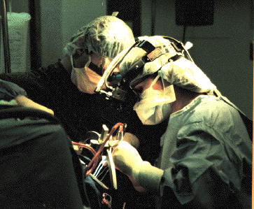
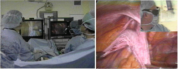
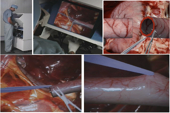
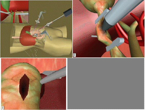
Endoscopy is minimally invasive surgery through natural body openings or small artificial incisions ('keyhole surgery'): laparoscopy, thoracoscopy, arthroscopy, and so on. A small endoscopic camera is used in combination with several long, thin, rigid instruments. The trend is to carry our as much surgery as is feasible by this means, to minimise the risk to patients.
Endoscopic surgery: the current situation without VR
Advantages for the patient include less pain, and less strain on the organism, and faster recovery. There are also relatively small injuries, and an economic gain arising through shorter illness time.
However, for the surgeon, there are several disadvantages, including restricted vision and mobility, difficult handling of the instruments, difficult hand-eye coordination and no tactile perception except force feedback.
Endoscopic surgery is becoming increasingly popular, because of its significant advantages. It is also the most popular surgical application of VR, partly because it expands on what is already an "unnatural" view of the locus of operation. Another reason is that endoscopic surgery is relatively easy to simulate because of the limited access, restricted feedback (especially tactile) and limited freedom of movement of instruments. Endoscopic simulators are being produced by all the main medical VR companies, usually with a focus on training.
Another recent trend is towards so-called Virtual Endoscopy. This is a technique whereby data from non-intrusive sources - such as scans - are combined into a virtual data model that can be explored by the surgeon as if an endoscope were inserted in the patient. VR is increasingly being used to provide surgeons with a meaningful and interactive 3D view of areas and structures they would otherwise be unable or unwilling to deal with directly.
In radiosurgery, X-ray beams from a Linear Accelerator are finely collimated and accurately aimed at a lesion. Popular products include Radionics X-knife, and Elekta`s Gammaknife.
Planning radiosurgery is suitable for VR, since it involves detailed understanding of 3D structure.
Elekta's Gammaknife(left ) and the X-knife from Radionics (right)
VR in surgery differs from most other VR in its focus on contact with objects, which must often be deformable objects and interdependent. The focus is on looking into objects rather than looking into space - there is less room available. The data is essentially volumetric and finger and hand interaction must be extremely precise.
The above characteristics bring with them certain technical requirements, such as real-time response to user`s action - which implies fast graphics, low latency input devices. The images must be of high resolution and faithful to the actual patient data, since life-critical decisions are based on the presentation of patient data. For simulators, the physical procedures must match those used in the actual operation.
Other requirements of VR for surgery include registration of patient data with atlases and the ability to coregister multimodal data. For use over extended periods, which is often needed in surgery, the style of user interaction should be natural, comfortable, and easy to use.
Areas where VR is being applied:
VR can in principle be applied to enhance reality for image-guided surgery. When applied to image-guided surgery in this way, the images obviously need to be available intra-operatively, and accurate registration of the real patient with the data becomes a crucial issue.
Currently, VR is used much more for preoperative planning (see 3.4 below) than to guide actual surgery (due to the understandable conservatism of medical practitioners). When VR is used intra-operatively, it tends to be implemented as some form of Augmented Reality (see the University of North Carolina system, below, and 2.4 above). Image-guided surgery is also a prerequisite of remote telemedicine and collaboration (see 3.5 below).
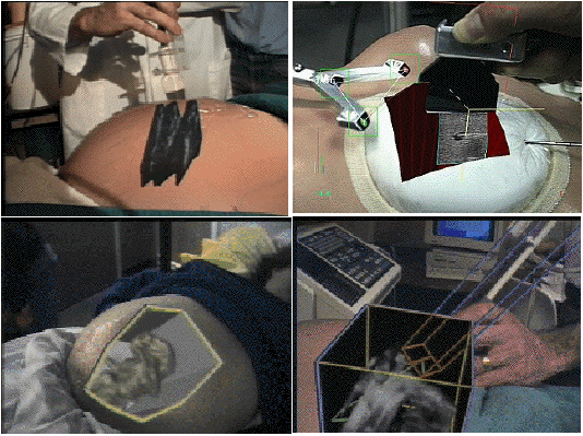
Image-guided surgery, implemented as Augmented Reality, at the University of N. Carolina
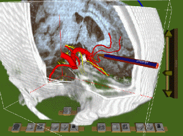
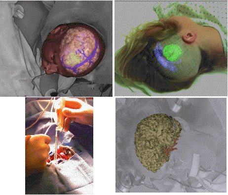
Brain tumour surgery guidance images
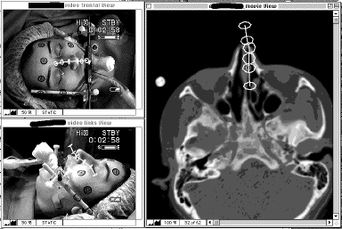
ARTMA (University of Vienna) system for Image-guided Ear, Nose & Throat Surgery
The ARTMA team at the University of Vienna were pioneers in this field. They refer to their approach as Interventional Video Tomography (see abstract below). It is also applied to telemedicine (see 3.5 below).
SPIE Proceedings of Lasers in Surgery, 4-6 February 1995, San Jose, CA
Paper #: 2395-34, pp.150-152
Author(s): Michael J. Truppe, Ferenc Pongracz, Artma Medizintechnik GmbH, Wien, Austria; Oliver Ploder, Arne Wagner, Rolf Ewers, Universität Wien, Vienna, Austria.
Abstract
Interventional Video Tomography (IVT) is a new imaging modality for Image Directed Surgery to visualize in real-time intraoperatively the spatial position of surgical instruments relative to the patient's anatomy. The video imaging detector is based on a special camera equipped with an optical viewing and lighting system and electronic 3D sensors. When combined with an endoscope it is used for examining the inside of cavities or hollow organs of the body from many different angles.
The surface topography of objects is reconstructed from a sequence of monocular video or endoscopic images. To increase accuracy and speed of the reconstruction the relative movement between objects and endoscope is continuously tracked by electronic sensors. The IVT image sequence represents a 4D data set in stereotactic space and contains image, surface topography and motion data. In ENT surgery an IVT image sequence of the planned and so far accessible surgical path is acquired prior to surgery. To simulate the surgical procedure the cross sectional imaging data is superimposed with the digitally stored IVT image sequence. During surgery the video sequence component of the IVT simulation is substituted by the live video source.
The IVT technology makes obsolete the use of 3D digitizing probes for the patient image coordinate transformation. The image fusion of medical imaging data with live video sources is the first practical use of augmented reality in medicine. During surgery a head-up display is used to overlay real-time reformatted cross sectional imaging data with the live video image.
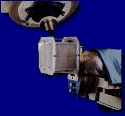
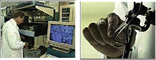
A main tool for traditional image-guided surgery is the microscope. Microscopes are now being integrated with robotic transport systems. Microscopes can also, in principle, serve as the vehicles for VR, as they will increasingly allow 3D views and are already "in place" in the operating theatre. Surgeons readily accept microscope-based views from which they can easily look away, whereas they are less comfortable with overlays placed on their primary direct view of the patient - the traditional augmented reality approach.
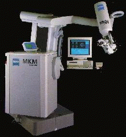
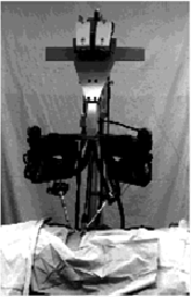
The Zeiss MKM Microscope transport. It is a 6 degree of freedom robot with a surgical stereomicroscope attached.
VR provides a unique resource for education about anatomical structure. One of the main problems for medical education in general is to provide a realistic sense of the inter-relation of anatomical structures in 3D space. With VR, the learner can repeatedly explore the structures of interest, take them apart, put them together, view them from almost any perspective. This is obviously impossible with a live patient, and is economically infeasible with cadavers (which, in any case, have already lost many of the important characteristics of live tissue).
Another advantage of VR for medical education is that demonstrations and exercises or explorations can easily be combined. For example, a "canned" tour of a particular structure, perhaps with voice annotations from an expert, can be used to provide an overview. The learner may then explore the structure freely and, perhaps later, be assigned the task of locating particular aspects of this structure. It is also possible to preserve particularly instructive cases, which would be impossible by other means.
There is something of crisis in current surgical training. As the techniques become more complicated, and more surgeons require longer training, fewer opportunities for such training exist.
Training in the operating theatre itself brings increased risk to the patient and longer operations. New surgical procedures require training by other doctors, who are usually busy with their own clinical work. It is difficult to train physicians in rural areas in new procedures. Training opportunities for surgeons are on a case-by-case basis. Animal experiments are expensive, and of course the anatomy is different.
The solution to these problems is seen to be the development of VR training simulators. These allow the surgeon to practice difficult procedures under computer control. The usual analogy made is with flight simulators, where trainee pilots gain many hours of experience before moving on to practice in a real cockpit.
Boston Dynamics open surgery anastomosis trainer
The advantages of training simulators are obvious. Training can be done anytime and anywhere the equipment is available. They make possible the reduction of operative risks associated with the use of new techniques, reducing surgical morbidity and mortality.
However, the big challenge is to simulate with sufficient fidelity for skills to be transferred from performing with the simulation to performing surgery on patients. Faithfulness is hard to achieve and much more evaluation of different approaches to training simulation are needed. Many experienced surgeons predict that in time, experience with training simulators will constitute a component of medical certification. But this will require new regulations and legislation.
Hot topics in the area include the use of force feedback (see 2.3.4 above), increased accuracy of modelling of soft tissue, and the role of auditory feedback.
For simple operations like suturing and biopsy needle placement, VR is effective, but perhaps an overkill to train skills that can easily and cheaply be acquired in other ways.
The most useful and tractable areas for the development of training simulators are the various techniques of endoscopic surgery in widespread use today. It is relatively easy to reproduce in VR the restricted field of view and limited tactile feedback of endoscopic surgery. It is much more problematic to reproduce open surgery techniques realistically. For complex anatomical structures, this is definitely not yet possible.
Karlsruhe Endoscopic Surgery Trainer
The pictures above illustrate both the value of simulators for training procedures, but also their current weaknesses in terms of realism. To realistically simulate an operation, the method of interaction should be the same as in the real case (as with flight simulators). When this is not the case, the VR can serve as an anatomy educational system rather than a training simulation.
One way of increasing the reality of interaction is to combine VR with physical models, as illustrated in the Gatech simulators for endoscopy and eye surgery, and the Penn State University bronchoscopy simulator (see below). These systems focus on training the surgeon in the use of particular medical devices, rather than on training a better awareness of general or specific patient anatomy.
Gatech: Endoscopic Surgical Simulator
Bronchoscope Simulator from Penn State University Hospital at Hershey
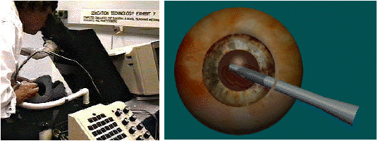
An example of an anatomy educational system is the EVL eye (shown below) from the University of Illinois. Since the VR is immersive and based around the CAVE, it cannot be said to duplicate the interaction methods of real eye surgery (since surgeons cannot get physically inside eyes) and so is not a training simulator, unlike the Georgia Tech system above.
The EVL eye, from the Electronic Visualisation Lab, University of Illinois at Chicago
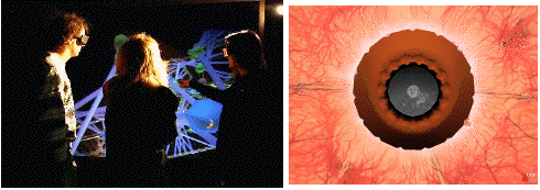
The EVL eye used by a group in the CAVE
More realistically in terms of interaction, the Responsive Workbench is another candidate for anatomy teaching (see below). As with CAVE-based applications, a shared VR enhances the potential for collaborative learning.
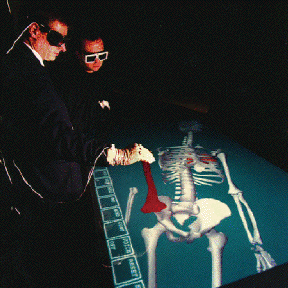
The Responsive Workbench from GMD in Germany
The most technologically challenging area of simulator training is for highly specialised aspects of life-critical operations such as brain surgery. The Johns Hopkins/KRDL skull-base surgery simulator for training aneurysm clipping (see below) is one example. The interaction is entirely with the VR itself.
JHU/KRDL Skull-base Surgery Simulator
Researchers at University of California San Diego Applied Technology Lab have developed an interesting Anatomy Lesson Prototype [http://cybermed.ucsd.edu/AT/AT-anat.html]. They point out that the main challenges they identified from talking to medical faculty and students included visualising potential spaces; studying relatively inaccessible areas; tracing layers and linings; establishing external landmarks for deep structures; and cogently presenting embryological origins. Correlating gross anatomy with various diagnostic imaging modalities, and portraying complex physiological processes using virtual representation were also considered highly valuable goals.
Relevant Web Sites:
Simulators such as the JHU/KRDL Skull-base Surgery Simulator blur into systems for pre-operative planning. Planning systems also sometimes blend with augmented reality, since the planning is on a actual, particular patient, so that physical reality (the patient) and the VR naturally come together in planning. The aim in such planning is to study patient data before surgery and so plan the best way to carry out that surgery.
Preoperative planning must:

Radionics' Stereoplan - a 'pure' planning system
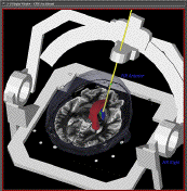
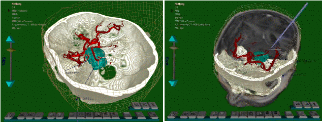
The aim of Stereoplan is to allow surgeons to examine patient data as fully as possible, and evaluate possible routes for intervention. Further, the system then provides the coordinates for the stereotactic frames that are standardly used to guide the route for brain surgery. Similar to the Radionics' Stereoplan, the KRDL Brainbench, built around the Virtual Workbench, aims at helping planning of stereotactic frame-based functional neurosurgery (see below).
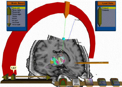
KRDL Brainbench for stereotactic frame-based neurosurgery planning
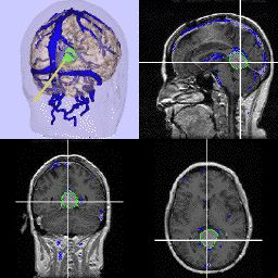
Combined neurosurgery planning and augmented reality from Harvard Medical School
In pre-operative planning the interaction method need not be realistic and generally is not. The main focus is on exploring the patient data as fully as possible, and evaluating possible intervention procedures against that data, not in reproducing the actual operation. The University of Virginia "Props" interface illustrates this (below). A doll's head is used in the interaction with the dataset, without any suggestion that the surgeon will ever interact with a patient's head in quite this way.
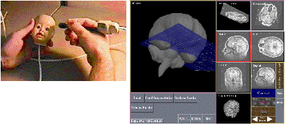
University if Virginia "Props" Interface used in pre-operative plannng
KRDL VIVIAN: the Virtual Workbench used for stereotactic tumor neurosurgery planning
Of course, the simulation must be accurate. Given this, techniques developed for planning can sometimes be applied to the prediction of outcomes of interventions, as in bone replacements or reconstructive plastic surgery. Such simulations can also help in training, and in communications between doctors and patients (and their families).
An important aspect of such systems for use by medical staff is the design of the tools and how this affects usability. See "Interaction Techniques for a Virtual Workspace".
Telemedicine is surprisingly little used today in actual medical practice. According to a recent article (The Economist, February 28th 1998) less than 1 in 1000 radiographs are viewed by a distant, rather than a local, specialist. This is despite the proven ability of telemedicine to save doctors' time and, hence, money (for example, from a recent study in Tromso on teleradiology). Similarly, home visits can be successfully replaced with remote consultations, saving money and increasing aged patients' satisfaction (because they can get more frequent consultations without troublesome travel), but currently only 1 in 2000 home visits are conducted remotely through information technology. Telemedicine is successfully used in military settings, where normal legal and economic considerations do not apply.
One promising area where VR could make a contribution is in remote diagnostics, where two surgeons can confer on a particular case, each experiencing the same 3D visualisation, although located in different places.
The other, often discussed, main applications are for remote operations, either through robotic surgery, or through assistance to another remote surgeon. The big problem here is network delay, since almost immediate interactivity is required. The small delay introduced by the use of satellite communication is unacceptable in remote surgery. Talk of remote operations carried out on space crew in deep space, or even merely on Mars, is pure science fiction (they would require communication at speeds above that of light).
Robots are used more routinely non-remotely, for precision in carrying out certain procedures, such as hip replacement. The types of operation to which robots are applied in this way are usually high volume, repeated procedures. As well as improved accuracy, major cost savings can be produced.
A relatively new development is to use surgeon-controlled robots to carry out, by key-hole methods, operations which previously required open surgery. VR becomes important here in providing a detailed 3D view to guide the surgeon in carrying out the operation via extremely small robotic instruments. Major operations, such as coronary bypass, can be carried out in this way with significantly reduced trauma and recovery time for the patient.
The technical possibility already exists for unsupervised robots to carry out surgery, but much ethical and legal debate and legislation will be needed before this could be put into practice. This survey does not focus primarily on telerobotics, which is itself a large field.
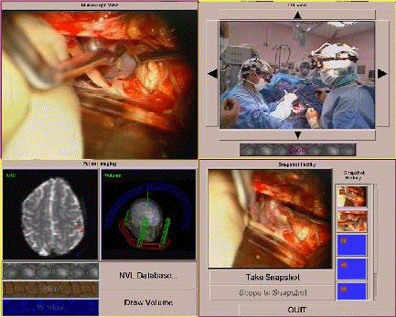
Remote surgeon workstation at the University of Virginia
The two upper images are live video provided via an ATM link. They show a view through the surgical microscope and a room view. The remote surgeon may pan/tilt/zoom the room camera and may move the microscope view with 6 degrees of freedom. The bottom windows respectively show presurgical imaging with functional overlays, volume rendering of the surgical plan, and a snapshot archive taken during surgery.
The Artma Virtual Patient System is an established technology for telesurgery.
The SRI Telepresence project is representative of current work in this area (see below).
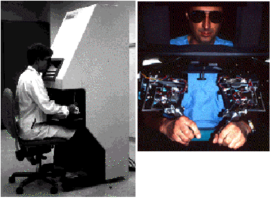
SRI Telepresence: telerobotics and stereo video interface, surgeon interaction
SRI Telepresence: telerobotics and stereo video interface, patient interaction
Much of the research in this area, especially in the USA, is funded by the government for military applications such as remote surgery on the battlefield. The main agencies re DARPA and Yale-NASA (mostly the same projects that previously got funding from DARPA). Progress reports on these projects, may of which have been running for several years, were presented at the conference and included:
During the conference as a whole, a broad selection of current work, both academic and commercial was presented. There were many endoscopic simulators, for the knee, shoulder, colon, abdomen... And all had some force-feedback that wasn't convincing as real tissue (from what doctors said) but apparently helped in training (from what the engineers said).
Tactile tissue simulation was one of the key phrases. Everybody is trying to figure out how to do it, but I didn't see (feel) any convincing implementation. Force feedback is the latest craze, but the sensitivity to model subtle gradations just isn't there yet. An interesting alternative is to use sound as feedback.
Also, many atlases of the whole human body (and one of a frog) were presented. Most used the Visible Human, but there were others (the Japanese) that had their own data sets.
One interesting point that was raised by the team at SRI is that the key problem in training surgeons is not how to convey the locomotive skills needed to manipulate an endoscope or how to cut using a scalpel, but how to understand patient anatomy. Training the hands how to use an endoscope takes a week or so, but learning how to the interpret a patient's anatomy takes years. I agree with this assessment, and I think that's where rich interaction capabilities combined with real-time volumetric rendering of multimodal data are crucial.
SRI, of Stanford, have tested their telepresence system with live animals using a 200 metre link. Their results are published in the Journal of Vascular Surgery. Dr. John Hill of SRI presented their first attempts to move towards computer-generated graphics training simulators using their telepresence system. They use a set-up similar to the ISS Virtual Workbench, but with their own interaction devices. They are working on simulating suture of tissue and vessels using an Onyx and 2D texture maps.
Dr. Ramin Shahidi, Stanford University Medical Center, is working on SGI-based volume rendering neurosurgery and craniofacial applications. Their graphics didn't include more than one volume at a time. His presentation was an overview of the use of volume rendering vs. surface rendering.
NASA-Ames and Stanford University have created the National Biocomputation Center: Dr. Muriel Ross was announcing this centre as a resource for collaboration with academics and industry, to promote medical VR. NASA-Ames have an Immersion Workbench (aka Responsive Workbench, aka Immersadesk) and their own visualisation software and are working on craniofacial "virtual" surgery. It appears that they use polygon meshes for their visualisation.
Dr. Henry Fuchs presented work in progress at UNC that uses depth range finders to reconstruct a surface map of the intestines to then guide an endoscope for colonoscopy. All this was added to their well-known augmented reality system, and comprises an interestingly novel approach.
HT Medical presented their VR Simulation of Abdominal Trauma Surgery. They use the PHANToM and some "wet" graphics to remove a kidney. They simulate the "steps" taken by the surgeon. First the surgeon cuts the skin, which then opens, revealing the intestines. A wet graphics effect is used, but this looks more like "cling film" wrapping over everything. The intestines moved quite unconvincingly, in an animation that was slightly under the control of the user (it didn't appear like inverse kinematics were attaching the end-point of the intestines to the user's tool). The kidney was removed by simply "reaching into it" and moving it out. The practical value of such a demonsytration was not clear, however.
An impressive paper from Wegner and Karron of Computer Aided Surgery Inc., described the use of auditory feedback to guide blind biopsy needle placement. Their audio feedback system generates an error signal in 3D space with respect to a planned needle trajectory. This error signal and the preoperative plan are used to motivate a position sonification algorithm which generates appropriate sounds to guide the operator in needle placement. To put it simply, harmonics versus dissonances are used to convey position information accurately along 6-8 dimensions. A nice example of a synaesthetic medium - using one modality (sound) where one would normally expect another (touch and/or vision). Their approach has wide applicability.
Myron Kreuger is President of Artificial Reality Corporation and a claimant to the title of inventor of VR. The system he described was a training system for dealing with emergencies, where smells of, for example, petrol or the contents of the lower intestine, can provide valuable information in a hazardous situation. It has also been claimed that smell can significantly enhance the effectiveness of medical training systems.
Many manufacturers presented their latest demonstrations and products at the conference exhibition. HT demonstrated CathSim, a simulator that trains nursing students to perform vascular catheterisations. They built a special force feedback device and a some simple graphics to provide visual feedback. It was quite good to guide the needle, but had little or no feedback once inside the skin. This seemed like an "technological overkill" since the procedure is easily learned without VR and is not exactly hazardous.
They also demonstrated a Flexible Bronchoscopy simulator developed with a partnership of pulmonologists and pharmacology experts at Merck & Co. (based on the Visible Human Project). They have a way to track the flexible tip of the endoscope ("a secret", I was told when I asked) and they generate nice 2D texture-mapped graphics of the interior throat using an SRI Impact.
Fraunhofer had two demonstrations from their Providence office:
Loral were at the Immersion booth, presenting a training system using the Immersion Corp.'s force feedback device. The application lets the surgeon guide an endoscope through the nose of a patient. The simulation was "helpful" to surgeons, although it is rather crude and doesn't feel like the real thing.
Prosolvia (the main Swedish VR company) demonstrated s system for Virtual Arthroscopy of the shoulder, developed with University Hospital of Linköping. They used the Immersion Corp. force-feedback system, and their own Oxygen software base. They are interested in collaborating on VR medical training systems.
Four demonstrations were shown at the SensAble booth:
Sense8's medical customers are the National Centre for Biocomputation (NASA, Stanford University), Rutgers University, Center for Neuro-Science, and Iowa School of Dentistry. A knee simulator was presented. Unfortunately, it broke early in the conference.
Vista Medical Technologies had a good head mounted display to substitute the microscope. It is not head tracked, but it allows the surgeon to look through the microscope and outside. It also allows Picture-in-Picture, so that an endoscope can be used to supplement the microscope.
There was a nice demonstration of 3D sonification from Lake Acoustics of Australia, who were also involved in the 3D sound feedback for biopsy needle placement described briefly above (the paper by Wegner and Karron). Using their kit, it is very simple to place sounds in a three-dimensional landscape surrounding the body to the front (as with normal stereo) and to the back (as with cinema surround sound) but using only headphones. They were giving away diskettes containing an impressive demonstration of this system.
It is clear that this is one of the medical areas where VR can most immediately and successfully be applied today. This is partly because the technical demands, particularly in terms of detailed visualisation and interactivity, are actually less stringent than in some other areas, such as surgery. Often these systems simulate the physical environment, a world of rooms, doors, buildings, etc., many of which are simple shapes and much easier to model that the irregular and contoured surfaces of internal organs. They also tend to be solid, and so the physics to be understood so that they may be modelled is much simpler, and the complexity of interacting with them is much less.
Main Application areas:
Examples:
3.7.1 Snapshot of the State of the Art: Conference Report on Mental Health session, Medicine Meets VR 1998
Topics covered at this year's conference included treatment of phobias, psychological assessment, and cognitive rehabilitation.
The session also provided an opportunity for the launch of the new CyberPsychology and Behavior journal, the first number of which includes a useful summary of the use of VR as a therapeutic tool.
Brenda Wiederhold presented a good paper on using VR to go beyond the standard "imaginal" training of phobic patients. The advantages of VR are, first, that fear can be effectively activated (which is necessary to bring about change) but can be controlled (too much fear reinforces the phobia) and, second, physiological measures can be used to control the display. One simple measure of anxiety, first used by Jung, is a drop in skin resistance.
Similar work on claustrophobia and fear of heights was described by Bulligen of the University of Basle. Another paper on acrophobia (fear of heights) by Huang et al. of the University of Michigan described comparisons of real and virtual environments for emotional desensitisation, and questioned the need for a high level of realism. Using the CAVE environment, they compared the same views in VR and in reality. See their Web page for views.
A rather pleasant system from Japan, the "Bedside Wellness" system by Ohsuga et al, allows bedridden patients to take a virtual forest walk while lying on their backs in bed. An array of three video screens present the unfolding view of the forest as the patient gently steps on two foot pedals. There is also 3D sound of birds, streams and wind in the trees. A slot below the central screen delivers a gentle breeze scented with pine to the "walking" patient.
Rizzo, of the University of Southern California, is using VR to give increased ecological validity to standard tests applied to Alzheimer's Disease patients, such as the mental rotation task (where the patient has to decide if a second figure is a rotated version of an earlier figure, or is different in shape). This Immersadesk application seemed like technological overkill to me. However, a fuller paper by Rizzo et al in the CyberPsychology and Behavior journal, lists several advantages of VR for cognitive and functional assessment and rehabilitation applications:
2. total control and consistency of administration
3. hierarchical and repetitive stimulus challenges that can be readily varied in complexity, depending on level of performance
4. provision of cueing stimuli or visualisation tactics to help successful performance in an errorless learning paradigm
5. immediate feedback of performance
6. ability to pause for discussion or instruction
7. option of self-guided exploration and independent testing and training
8. modification of sensory presentations and response requirements based on user's impairments
9. complete performance recording
10. more naturalistic and intuitive performance record for review and analysis by the user
11. safe environment, although realistic
12. ability to introduce game-like aspects to enhance motivation for learning
13. low-cost functional training environments
Joan McComas of the University of Ottawa described a VR system for developing spatial skills in children. She had carried out a four-condition study where choice of location to move to was either passive or active, as was navigation to that location. The four were then: passenger (passive choice/passive movement) navigator (active choice/passive movement), driver (active choice/active movement) and navigated driver (passive choice/active movement). The task was to find things hidden at locations, but without going to the same location twice. Measures were percent of correct choices and visit of first error. It occurred to me that we could use this sort of approach in studies of exploration in 3D information landscapes. A paper by Weniger also struck a chord by comparing spatial learning (maze navigation) with exercise of the executive function (the maze with pictograms) and with the use of orientation skills (navigation of landscapes).
Giuseppe Riva, from the Applied Technology for Psychology Lab at the Instituto Auxologico Italiano in Verbania also discussed the use of VR for psychological assessment - particularly the development of the Body Image Virtual Reality Scale. Patients chose which virtual body they think matches their own, and which they would prefer to have instead. The difference gives a measure of body image distortions.
Greene and Heeter, of the Michigan State University Communication Technology Lab, described CD-ROMs that contain VR-like stories of cancer sufferers, particularly in relation to coping with pain. Details can be found at [http://www.commtechlab.msu.edu/products/]. An interesting paper by Hunter reported the finding that VR can be very effective in helping burn-recovery patients cope with the pain of treatment. Patients in the VR condition reported significant pain reduction and less time spent thinking about pain.
Pope described the use of a VR system called "Viscereal" to provide physiological feedback. Users could control the flow of blood to their hands, and hence could warm or cool them at will. It has also been found to be effective in permitting conscious control of bowel activity, easing clinically harmless but distressing conditions such as Irritable Bowel Syndrome.
The Woodburys, a husband and wife team from the Puerto Rican Institute of Psychiatry, mused on modern cosmology and the origins of our three dimensionality. They gave the conference a useful reminder that the 3D world is in our heads, not in the world "out there". Pathological psychological states - especially various psychoses - and altered states of consciousness produced by certain hallucinogenic drugs, make this clear as the world around the experiencer, and his sense of his body and its place in that world, falls apart in typical psychotic panic states. Following Pribram, the Woodburys view the 3D world we know so well as a holographic projection, formed in the brain according to principles established through evolution as aiding survival. While recognising that this world is an illusion, psychiatrists work to restore it in patients whose world has literally collapsed.
Although not mentioned by presenters, one of the audience, Rita Addison, talked about the use of VR to communicate the reality of mental deficits to other, normal people. Rita has visited the VRLab in Umea and is well-known for her "Detour: Brain Deconstruction Ahead" which reproduces for others her visual problems since a car accident a few years ago.